651 Views
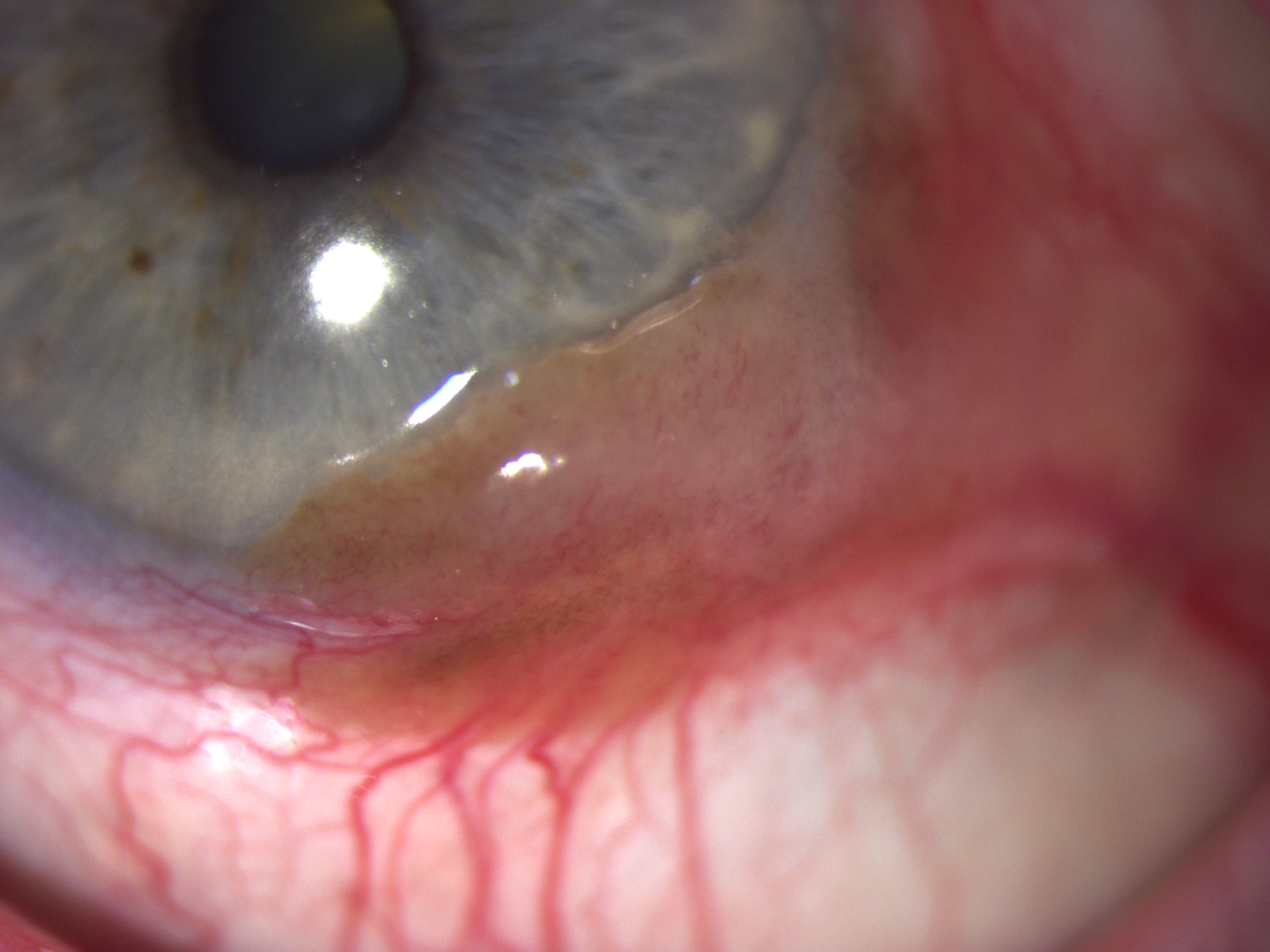
Pigmented Limbal Lesion
This 85 caucasian female presented with a large left temporal limbal lesion measuring 9 x 3 mm. There was mild pigmentation to large feeder vessels and adjacent PAM. Initial topical treatment with Mitomycin C for chemoreduction was commenced and the lesion excised with cryotherapy. Histology showed an amelanotic melanoma. MRI and LFT were clear.